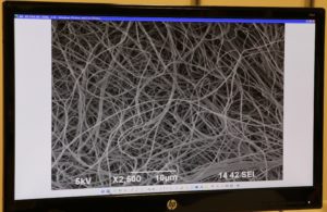
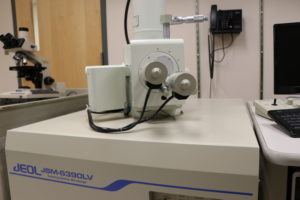
 The Indiana University School of Medicine Electron Microscopy (EM) Center is a resource to help investigators look at specimens at a higher magnification than they would be able to with a regular microscope. The lab offers both transmission electron microscopy and scanning electron microscopy, with the ability to magnify specimens up to 300,000 times the original size.
 “Most of the people who come to the EM core are looking at morphological changes in their tissue specimens that they can’t see at the light level,” said Caroline Miller, who is the core’s assistant director.
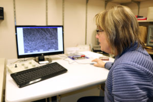
Miller manages the IU School of Medicine EM Center, which includes determining if projects are suitable for electron microscopy, deciding which piece of equipment to use for the experiment and preparing specimens for viewing on the microscopes. She says they look at both biological and material specimens as samples.
“I never know what’s going to come through my door,” said Miller. “When I can help researchers get to a point where they’ve figured something out, to help with their grant or just get to the next step in their research, that makes it fun.”

Miller says the Indiana Clinical and Translational Sciences Institute (CTSI) has helped several investigators lay the groundwork of their research in the EM lab by providing grants, and then those initial results have led to external funding for researchers. In the 50 years Miller has worked in the EM field, she says technology has changed, but also stayed the same.
“The electron microscopes have gotten better,” said Miller. “They’re easier to use. I used to have to take film out and develop it, then print the pictures off from the negatives. Now I can just send digital images via Box to all my researchers. But there are a lot of things that are still very basic to utilizing a microscope. The limitations that are involved will never change.”
Any Indiana CTSI investigator can use the EM core facility. For more information, click here.
